
The superior portion of the vestibular nerve innervates three structures- the superior and lateral semicircular canals, and the utricle. The vestibular portion is further segmented into the superior and inferior portions at the fundus. In the lateral segment of the internal auditory canal, about 3 to -4mm from the fundus, the cochlear and vestibular nerves join to form one common nerve. The vestibulocochlear nerve runs most often posteriorly to the facial nerve in the internal auditory canal.
#Auditory meatus series
The posterior portion of the fundus is filled with a macula crista, which is a series of very small openings that the vestibular nerves pass to reach the superior and inferior semicircular canals. These two spines form three distinct osseous structures through which the facial and vestibulocochlear nerve branches can be found in a consistent pattern, represented by figure 1. The superior half is further divided into anterior and inferior segments by Bill’s bar, a vertical crest of bone named after otologist Dr. It is divided into superior and inferior segments by the transverse crest (also called the falciform crest). The fundus separates the internal auditory canal from the cochlea and vestibule which are located in close proximity. The canal narrows as it moves towards the fundus, a thin cribriform plate of bone that marks the lateral boundary of the canal. The rounded and smooth canal is on average 8.5mm (5.5 to 10.mm) in length and about 4mm in diameter. It is lined by dura and filled with spinal fluid. The canal runs through the petrous segment of the temporal bone, which is located between the inner ear and posterior cranial fossa. The internal auditory canal begins in the temporal bone within the cranial cavity at an oval-shaped opening called the porus acusticus internus. Knowledge of the anatomy and relationship of these structures plays a vital role during the evaluation and management of diseases involving the internal auditory canal. It includes the vestibulocochlear nerve (CN VIII), facial nerve (CN VII), the labyrinthine artery, and the vestibular ganglion. The tympanic membrane, is anatomically part of, and represents, the most medial extent of the external ear.The internal auditory canal (IAC), also referred to as the internal acoustic meatus lies in the temporal bone and exists between the inner ear and posterior cranial fossa.
#Auditory meatus skin
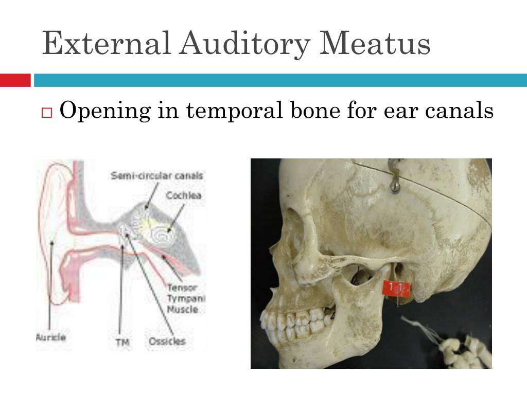
It has an outer cover of extremely thin skin and an inner layer of cuboidal epithelium facing the tympanic cavity. The tympanic membrane (or tympanum) consists of two layers of collagen fibers: It is approximately 3 cm long and is lined by skin containing hair follicles (tragi), sebaceous glands, and ceruminous glands (which produce cerumen). The external auditory meatus is a short S-shaped canal within the tympanic temporal bone leading from the external acoustic pore of the auricle to the tympanic membrane. ear lobe ( lobule): the lowest part of the ear and the only part that does not contain cartilage, situated below the intertragic incisure.cymba conchae: depression surrounded by the crus of the helix below and the inferior crus of antihelix above.cavum conchae: the deepest depression in the auricle, inferior to the crus of the helix.intertragic incisure: a notch separating the tragus from the antitragus.antitragus: situated in the lower part of the antihelix and faces the tragus.can be manually pushed back over the pore, to mitigate noise.tragus: prominence in front of the external acoustic pore.scaphoid fossa / scapha: the depression between the helix and antihelix.fossa triangularis: tiny depression between the crura.crura antihelicis: a pair of limbs located above the external acoustic pore.antihelix: a ridge parallel to the helix.crus helicis: anterior terminal portion of the helix superior to the external acoustic pore.
#Auditory meatus free


 0 kommentar(er)
